HAND & WRIST
The following experienced and skilled OrthoSouth® surgeons have a combined Google rating of
4.86 out of
5 based on
1614 ratings:


To view additional board-certified orthopedic physicians who are qualified to handle a range of hand and wrist issues, visit our provider page.
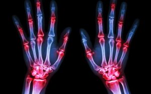
Anatomy of the Hand
Common Hand and Wrist Conditions
Carpal tunnel syndrome is the most common time of nerve compression in the hand and wrist area, affecting up to 10% of the population.
Causes/Risk Factors
- Female
- Obesity
- Pregnancy
- Hypothyroid
- Inflammatory Arthritis
- Elderly
- Kidney Failure
- Tobacco
- Alcoholism
- Repetitive Motion
- Mucopolysaccharidosis
- Mucolipidosis
- Vibration
- Heat/Cold Extremes
- Masses
- Diabetes
- Fractures
- Amyloidosis
Signs/Symptoms
- Numbness and tingling to the thumb, index, middle and/or ring fingers
- Pain and numbness at night
- Weakness to the hand
- Sensitivity to cold and hot temperatures
Diagnosis
A nerve study is a reasonable study to perform for several reasons. The nerve study helps in determining that the patient has carpal tunnel syndrome, how severe the nerve and muscle are affected, and the relative success that may be obtained following non-operative or surgical treatment.
Treatment
Non-Operative Treatment
Nonsteroidal anti-inflammatory medications may provide some symptoms relief. Bracing at least at night but potentially during the day may be effective treatment by decreasing pressure to the nerve. Activity modifications may be performed to reduce additional pressure to the nerve. Steroid injection may also be an alternative option for patients with carpal tunnel syndrome.
Surgery
Carpal tunnel release is performed to take pressure off of the nerve. Surgery may be performed open or endoscopically, which both give similar long term results for carpal tunnel treatment. Tendon transfers may be an additional surgery to consider if severe muscle weakness is present.
Causes
- Ganglion cyst
- Lipoma
- Repetitive Trauma
- Ulnar artery thrombosis/aneurysm
- Hook of hamate fracture
- Pisiform dislocation
- Inflammatory Arthritis
- Anatomic anomaly
Signs/Symptoms
- Pain and numbness to inner hand and small/wring finger
- Weak hand grip
Testing
A nerve study is a reasonable study to perform for several reasons. The nerve study helps in determining that the patient has ulnar nerve compression, how severe the nerve and muscle are affected, and the relative success that may be obtained following non-operative or surgical treatment.
Treatment
Non-Operative Treatment
Nonsteroidal anti-inflammatories may help in reducing symptoms of nerve compression as well as avoiding activities that worsen the symptoms. Patients may find wrist bracing helpful in treatment of ulnar nerve compression at the wrist.
Surgery
Release of tissue compression the ulnar nerve without or without release of the carpal tunnel is performed to provide relief of symptoms for ulnar nerve compression at the wrist. Tendon transfers may be an additional surgery if the patient presents with severe symptoms of clawing of the hand.
Trigger finger/trigger thumb is the loss of smooth tendon gliding within the A1 pulley of the finger and or thumb. It causes pain, clicking, catching, or ultimately locking of the finger. This condition affects up to 3% of the population and up to 10% of diabetics.
Risks Factors
- Female
- Diabetes
- Middle to Elderly Age Groups
- Inflammatory Arthritis
- Gout
- Pseudogout
- Hypothyroidism
- Sarcoidosis
- Amyloidosis
Symptoms
- Pain at base of finger or thumb at palm
- Mechanical symptoms of triggering of the finger or thumb
Treatment
Non-Operative Treatment
Nonsteroidal anti-inflammatory medications and activity modifications may provide some relief. Steroid injection(s) are typically reasonable options in most patients. Bracing may be an alternative option for patients with joint stiffness.
Surgery
Release of the pulley or tunnel usually provides relief for most patients. Some patients may possibly need excision of part of the flexor tendon for treatment.
Formally known as distal radius fracture, this injury is very common and represents up to 15-20% of all fractures in adults.
Risks
- Female
- Young and older populations
- Osteoporosis
Symptoms
- Pain and swelling at wrist
- Deformity at wrist
- Limited wrist motion
- Skin bruising and abrasion/laceration
Tests
X-rays and possible CT are usually obtained to diagnose a broken or fractured wrist.
Treatment
The goal of treatment is to have the broken wrist heal in good position and gain back functional motion to the hand, wrist, and forearm.
Non-Operative Treatment
Fractures that are not significantly displaced may be treated without surgery. Splints and casts are typically used but bracing may also be an option. The majority of distal radius and ulna fractures heal in 6 weeks.
Surgery
Fixation using pins called kwires, plates and screws, and possible exteneral fixators may be used depending on the type of fracture present.
- Closed reduction and pinning: may be performed with temporary pins that are removed once the fracture is healed.
- Open reduction and fixation: the surgeon makes an incision and directly visualizes the fracture. The bones pieces are then placed and held in good position by either pins or a plate(s) and screws.
- External fixation: Rarely, external pins are placed that are connected to bar(s) outside the skin. This method of fixation is usually reserved for severely open fractures and unstable patient’s in trauma setting.
Dupuytren’s contracture is an abnormal thickening of the tissue just beneath the skin. This thickening occurs in the palm and can extend into the fingers. Firm pits, bumps and cords (thick lines) can develop and cause the fingers to bend into the palm. This condition may also be known as Dupuytren’s Disease. Occasionally, the disease will cause thickening on top of the knuckles or cause lumps and cords on the soles of the feet (plantar fibromatosis).
Causes
Signs and Symptoms
Symptoms of Dupuytren’s contractureusually include lumps and pits within the palm. The lumps are generally firm and stuck to the skin. Thick cords may develop from the palm into one or more fingers. The ring and small fingers are most commonly involved. These cords may cause bending of the fingers. In many cases, both hands are affected, but each hand can be affected differently.
The lumps can be uncomfortable in some people, but Dupuytren’s contractureis not typically painful. The disease may first be noticed because of difficulty placing the hand flat on a surface. As the fingers are drawn into the palm, it may be more difficult to wash hands, wear gloves, shake hands, and get hands into pockets. It is difficult to predict how the disease will progress. Some people have only small lumps or cords while others will develop severely bent fingers. The disease tends to be more severe if it occurs at an earlier age.
Treatment
In mild cases, especially if hand function is good, only observation is needed. A lump in the palm does not mean that treatment is required or that the disease will progress. For more severe cases, various treatment options are available to straighten the finger(s). These options may include needles, injectable medicine or surgery.
Your hand surgeon will discuss the most appropriate method based upon the stage and pattern of the disease and the joints involved. The goal of treatment is to improve finger motion and function; however, complete correction of the finger(s) may not always happen. Even with treatment, the disease may come back. Before treatment, the surgeon will discuss realistic goals and possible risks.
Splinting and hand therapy are often needed after treatment in order to maintain the improved finger function.
This information is provided directly from the American Society for Surgery of the Hand.
Causes
A ganglion cyst can occur in patients of all ages. While their cause is unknown, the cysts may form in the presence of joint or tendon irritation, arthritis, mechanical changes, or injury.Signs and Symptoms
Your ganglion cyst may or may not be painful. The cysts are typically oval or round and may be soft or very firm. Cysts at the base of the finger on the palm side are typically very firm, smaller than a pea-sized nodule, and are tender to applied pressure, such as when gripping. Cysts at the far joint of the finger frequently have arthritis associated with them. At this end knuckle, the overlaying skin may become thin, and there may be a lengthwise groove in the fingernail just beyond the cyst.Diagnosing a Ganglion Cyst
The diagnosis is usually based on the location of the lump and its appearance. Light will often pass through these lumps, and this may assist in the diagnosis. Your physician may request x-rays in order to look for evidence of problems in adjacent joints.Treatment
Treatment for a ganglion cyst can often be non-surgical. In many cases, these cysts can simply be observed, especially if they are painless. Ganglion cysts frequently disappear spontaneously. If the cyst becomes painful, limits activity, or is otherwise unacceptable, several treatment options are available, including:- Splints and anti-inflammatory medication to decrease pain associated with activities
- Aspiration to remove the fluid from the cyst and decompress it (This requires placing a needle into the cyst, which can be performed in most office settings. Aspiration is a very simple procedure, but recurrence of the cyst is common.)
If non-surgical options fail to provide relief, or if the cyst reoccurs, surgical alternatives are available. Surgery involves removing the cyst and may include removal of a portion of the joint capsule or tendon sheath. In the case of wrist ganglion cysts, both traditional open and arthroscopic techniques usually yield good results. Surgical treatment is generally successful, although cysts may reoccur. Your hand surgeon will discuss the best treatment options for you.
This information is provided directly from the American Society for Surgery of the Hand.
A joint is formed when two bones meet and articulate, which allows movement. Joints are lined with smooth cartilage that allows for the easy movement of one bone relative to another. Osteoarthritis, or degenerative arthritis, is a process that occurs with aging and describes the deterioration of the joint cartilage. Thumb arthritis is the second most common type of arthritis in the hand; the most prevalent hand arthritis involves the last joint in each finger.
Thumb arthritis is also known as basal joint arthritis. It is more common in women, though certainly men can develop this type of problem. Typically, degenerative arthritis of the thumb occurs sometime after 40 years of age. There is a genetic predisposition in developing this arthritis condition. Additionally, any type of trauma to the thumb can predispose one to thumb arthritis. There are other conditions such as inflammatory arthritis (Lupus, Rheumatoid Arthritis) that can affect the base of the thumb and cause the deterioration of that joint.
Signs and Symptoms
Activities which rely on the thumb may result in pain at the base of the thumb, particularly in an arthritic joint. The activities most people notice as painful include pinching, grasping or gripping. Signs and symptoms of thumb arthritis may include swelling or stiffness at the base of the thumb. As the arthritis progresses, the pain and weakness may increase along with decreasing range of motion at the base of the thumb. As the arthritis advances even further, bone spurs may develop, resulting in an enlarged appearance at the base of the thumb.
Diagnosis
If the pain starts to interfere with one’s activities of daily living, then a visit with a hand surgeon could be helpful. During a physical examination of an arthritic thumb, the physician may notice grinding when maneuvering the thumb in a certain way (see Figure 1). X-rays can be used to confirm the diagnosis.
Treatment
Treatment options are based on the severity of the symptoms. There is no convincing evidence to support the use of any medications or supplements to prevent the progression of thumb arthritis. X-rays show that basal joint arthritis will generally get worse with time. The rate of the progression of arthritis varies from one person to another. Even though thumb arthritis will advance with time, the symptoms do not always get worse, and in some people the symptoms will decrease significantly.
Nonoperative treatment for thumb arthritis can involve:
- Anti-inflammatory medications (consult your doctor first)
- The use of heat or ice
- Bracing
- Exercises
- Ergonomic adjustments
- Avoidance of irritating activities
- Steroid injections into the basal joint
- Occupational therapy
When nonoperative treatment fails and when the patient is sufficiently symptomatic, surgery is an option. Surgery can involve removing part or all of the trapezium (one of the bones in the thumb joint) and cushioning or suspending the thumb joint with a variety of possible techniques. Fusing (making the two bones into one) the thumb joint is also an option. The complete recovery after a thumb surgery can take anywhere between 8 weeks and one year.
This information is provided directly from the American Society for Surgery of the Hand.
The brachial plexus is a group of nerves that come from the spinal cord in the neck and travel down the arm. These nerves control the muscles of the shoulder, elbow, wrist and hand, as well as provide feeling in the arm. Some brachial plexus injuries are minor and will completely recover in several weeks. Other injuries are severe enough and could cause some permanent disability in the arm.
Causes
These nerves can be damaged by stretching, pressure or cutting. Stretching can occur when the head and neck are forced away from the shoulder, such as during a motorcycle fall or car accident. If severe enough, the nerves can tear out of the spinal cord in the neck. Pressure could occur from the crushing of the brachial plexus between the collarbone and first rib, which can happen during a fracture or dislocation. Swelling in this area from excessive bleeding or injured soft tissues can also cause an injury.
Signs and Symptoms
Nerve injuries can stop signals to and from the brain, preventing the muscles of the arm and hand from working properly, and causing loss of feeling in the area.
Treatment
Many adult injuries will not recover on their own, and early evaluation by physicians who have experience treating these problems is essential. Some injuries can recover with time and therapy. The time for recovery can be weeks or months. When an injury is unlikely to improve, several surgical techniques can be used to improve the recovery.
To help decide which injuries are likely to recover, your physician will rely upon multiple examinations of the arm and hand to check the strength of muscles and presence of feeling in different areas. Additional testing, such as an MRI scan or CT scan/myelography, may be used. A Nerve Conduction Study/Electromyogram (NCS/EMG), a test that measures the electrical activity transmitted by nerves and muscles, may also be performed.
In some cases, when nerve recovery will not happen, a tendon transfer surgery may be performed.
Recovery
The patient must do several things to keep up muscle activity and prevent the joints from getting stiff. Your doctor may recommend therapy to keep these joints flexible. If the joints become stiff, they will not move even after muscles begin to work again, like a hinge that has rusted.
When a sensory nerve has been injured, the patient must be extra careful not to burn or cut fingers while there is no feeling in the affected area. During nerve recovery, the brain may not interpret the new nerve signals properly, and a procedure called sensory re-education may be needed to optimize muscle control and feeling in the hand or fingers. Your doctor will recommend the appropriate therapy based on the nature of your injury.
Factors that may affect results after a brachial plexus injury include patient age and the type, severity and location of the injury. Though these injuries will result in lasting problems for the patient, care by a hand surgeon and proper therapy can maximize function.
This information is provided directly from the American Society for Surgery of the Hand.
The scaphoid is one of eight small bones that make up the “carpal bones” of the wrist. It connects two rows of these bones – the proximal row (closer to the forearm) and the distal row (closer to the hand). This connection puts it at extra risk for injury.
Causes
The scaphoid can be injured when a significant load is placed on the extended wrist, such as a fall onto an outstretched hand.
Signs and Symptoms
Most people with a scaphoid fracture (which is the same as a broken wrist) will have pain and/or swelling along the thumb side of the wrist within days following a fall. Because there is no visible deformity and no difficulty with motion, many people with this injury assume that it is a wrist sprain. Unfortunately, delaying treatment can cause problems. Visit a hand surgeon as soon as possible if you think you might have fractured your wrist.
Diagnosis
A scaphoid fracture is usually diagnosed by an x-ray of the wrist. However, x-rays do not always show scaphoid fractures. A break in the bone that cannot be seen on x-ray yet is called an “occult” fracture. If you are tender directly over the scaphoid bone (which is located in the hollow at the thumb side of the wrist as shown in Figure 2), your healthcare provider might recommend wearing a splint to be safe. If pain persists, a follow-up exam and x-ray in a week or two can be used to diagnose.
Sometimes, a CT scan and/or MRI is used to get better views of the shape and alignment of the scaphoid and assist with the diagnosis or surgery plans.
Treatment
If the scaphoid is broken, the few tiny blood vessels that supply the bone with nutrients can be damaged. Because blood supply is needed to heal a fracture, the scaphoid often takes a long time (a few months) to heal.
If the scaphoid fracture is non-displaced (bone has not moved out of place at the fracture), it usually can be successfully treated with a cast. Although the fracture may heal in as little as six weeks, it may take longer for some patients.
If the fracture is in a certain part of the bone or if the fracture is at all displaced (bone ends have shifted), surgery might be the best option. This might include the insertion of a screw or pins (Figure 3).
Scaphoid Non-Union Fractures
A scaphoid non-union fracture refers to a wrist fracture that is failing to heal. A fracture that is healing more slowly than expected is a “delayed union” fracture. If the scaphoid fracture is not healing, you may or may not continue to have symptoms. It may be a sign that your fracture is not healing if you notice decreased strength, such as inability to do push-ups.
Treatment of a scaphoid non-union fracture depends on many factors, including:
- Patient age
- Use of the hand
- Activity level
- Symptoms
- Cigarettes or other tobacco use
- Other medical and/or wrist conditions
- Fracture pattern
The goals of treatment are to relieve pain, maximize function and prevent arthritis. Usually, surgery is needed to clean out the fracture site, to potentially place some form of bone graft to help bone healing, and to stabilize the fracture with pins or screws.
Treatment without surgery can include use of a brace, anti-inflammatory medication or a cortisone shot for pain relief. You and your hand surgeon can discuss which treatment is best for you.
This information is provided directly from the American Society for Surgery of the Hand.
Additional Information